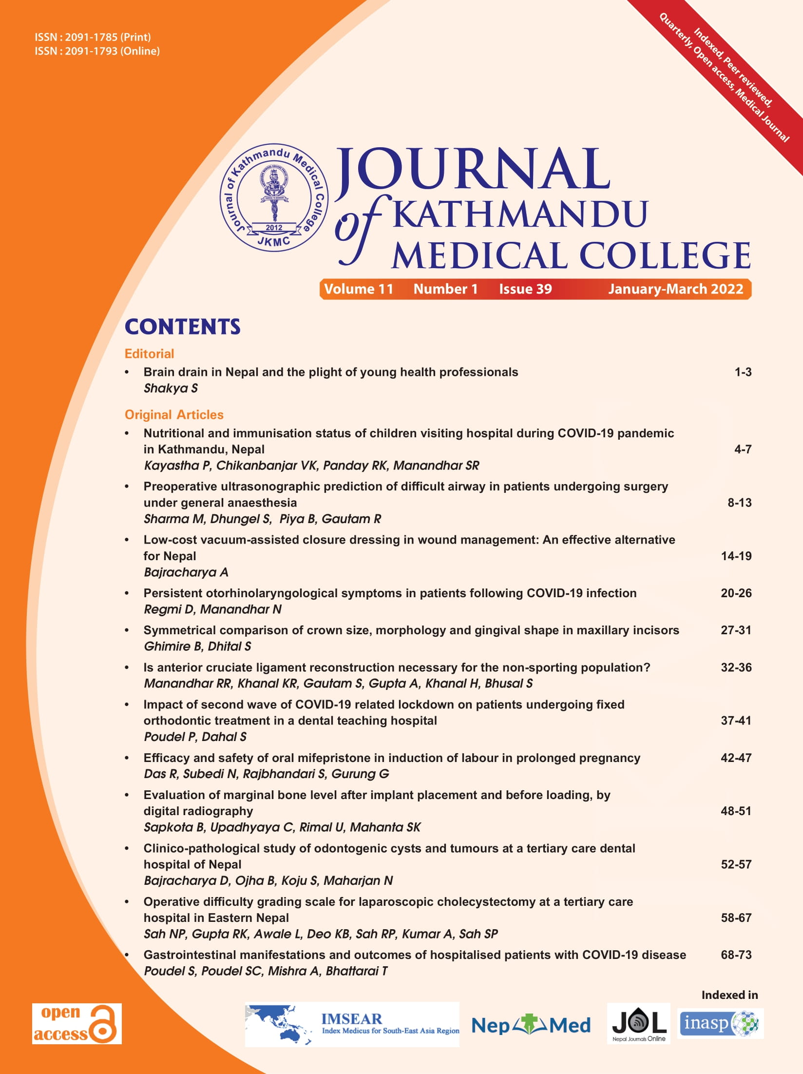
Evaluation of marginal bone level after implant placement and before loading, by digital radiography
DOI:
https://doi.org/10.3126/jkmc.v11i1.45494Keywords:
Bone loss, Distal, Implant, Mesial, OsseointegrationAbstract
Background: Marginal bone loss around dental implant is one of the criteria for evaluating implant success.
Objectives: To evaluate the marginal bone loss around dental implants using digital radiographs and to determine correlation of mesial and distal bone loss around dental implants with gender and location in either arch in a period of 3-6 months after placement and before prosthetic loading.
Methods: An analytical study was undertaken from July 2021 till December 2021, after ethical clearance, in 18 patients on whom 23 implants had been placed at Dhulikhel hospital. After implant placement first radiograph was taken by CE 0297 size 2 PSP plate and Carestream (CS2100) intraoral periapical radiograph machine using paralleling technique (XcpRinn Device) and second at 3-6 months later. The radiographs were viewed using image viewer software (Vistasoft2.0.1) to calculate the bone level. Calculating the difference in bone level at zero month and at 3-6 months gave us the amount of bone loss which was entered in Excel sheet and transferred to SPSS v.22 for analysis and student unpaired-t test was used.
Results: The mean bone loss was 0.27 ± 0.2 mm on mesial aspect and 0.13 ± 0.3 mm on distal aspect at the end of the study period. No statistically significant bone loss in relation to gender and location of implant placement was found.
Conclusion: Within the limitation of the study, no significant difference was found in the mesial and distal aspect of bone loss around dental implant when compared with different parameters.
Downloads
References
Zimmermann J, Sommer M, Grize L, Stubinger S. Marginal bone loss one year after implantation: A systematic review for fixed and removable restorations. Clin Cosmet Investig Dent. 2019 Jul 16;11:195-218. [PubMed | Full Text | DOI]
Szyma?ska J, Szpak P. Marginal bone loss around dental implants with various types of implantabutment connection in the same patient. J Preclin Clin Res. 2017;11(1):30-4. [Full Text | DOI]
Nandal S, Ghalaut P, Shekhawat H. A radiological evaluation of marginal bone around dental implants: An in vivo study. Natl J Maxillofac Surg. 2014;5(2):126-37. [PubMed | Full Text | DOI]
Koller CD, Pereira-Cenci T, Boscato N. Parameters associated with marginal bone loss around implant after prosthetic loading. Braz Dent J. 2016 MayJun;27(3):292-7. [PubMed | Full Text | DOI]
Lombardi T, Berton F, Salgarello S, Barbalonga E, Rapani A, Piovesana F, et al. Factors influencing early marginal bone loss around dental implants positioned subcrestally: A multicentre prospective clinical study. J Clin Med. 2019 Aug 4;8(8):1168. [PubMed | Full Text | DOI]
Shen XT, Li JY, Luo X, Feng Y, Gai LT, He FM. Peri-implant marginal bone changes with implant-supported metal-ceramic or monolithic zirconia single crowns: A retrospective clinical study of 1 to 5 years. J Prosthet Dent. 2021 Feb;S0022-3913(20)30800-3. [PubMed | Full Text | DOI]
Smith DE, Zarb GA. Criteria for success of osseointegrated endosseous implants. J Prosthet Dent. 1989 Nov;62(5):567-72. [PubMed | Full Text | DOI]
Bittar-Cortez JA, Passeri LA, de Almeida SM, HaiterNeto F. Comparison of peri-implant bone level assessment in digitized conventional radiographs and digital subtraction images. Dentomaxillofac Radiol. 2006 Jul;35(4):258-62. [PubMed | Full Text | DOI]
Singh P, Garge HG, Parmar VS, Viiswambaran M, Goswami MM. Evaluation of implant stability and crestal bone loss around the implant prior to prosthetic loading: A six month study. J Indian Prosthodont Soc. 2006;6(1):33-7. [Full Text]
Türk AG, Ulusoy M, Toksavul S, Güneri P, Koca H. Marginal bone loss of two implant systems with three different superstructure materials: A randomised clinical trial. J Oral Rehabil. 2013 Jun; 40(6):457-63. [PubMed | Full Text | DOI]
Bhardwaj I, Bhushan A, Baiju CS, Bali S, Joshi V. Evaluation of peri-implant soft tissue and bone levels around early loaded implant in restoring single missing tooth: A clinico-radiographic study. J Indian Soc Periodontol. 2016 Jan-Feb;20(1):36-41. [PubMed | Full Text | DOI]
Niu W, Wang P, Zhu S, Liu Z, Ji P. Marginal bone loss around dental implants with and without microthreads in the neck: A systematic review and meta-analysis. J Prosthet Dent. 2017 Jan; 117(1): 34-40. [PubMed | Full Text | DOI]
Galindo-Moreno P, Concha-Jeronimo A, LopezChaichio L, Rodriguez-Alvarez R, SanchezFernandez E, Padial-Molina M. Marginal bone loss around implants with internal hexagonal and internal conical connections: A 12-month randomised pilot study. J Clin Med. 2021 Nov;10(22):5427. [PubMed | Full Text | DOI]
Downloads
Published
How to Cite
Issue
Section
License
Copyright © Journal of Kathmandu Medical College
The ideas and opinions expressed by authors or articles summarized, quoted, or published in full text in this journal represent only the opinions of the authors and do not necessarily reflect the official policy of Journal of Kathmandu Medical College or the institute with which the author(s) is/are affiliated, unless so specified.
Authors convey all copyright ownership, including any and all rights incidental thereto, exclusively to JKMC, in the event that such work is published by JKMC. JKMC shall own the work, including 1) copyright; 2) the right to grant permission to republish the article in whole or in part, with or without fee; 3) the right to produce preprints or reprints and translate into languages other than English for sale or free distribution; and 4) the right to republish the work in a collection of articles in any other mechanical or electronic format.


