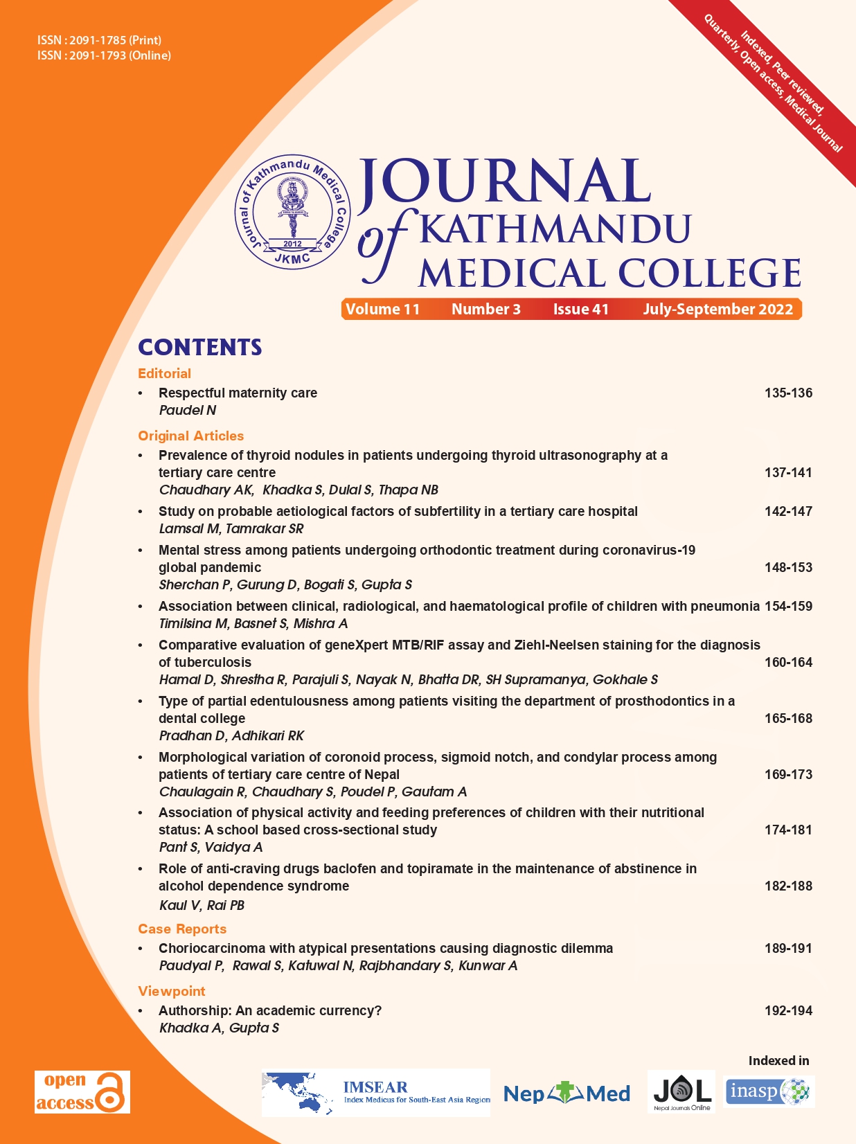
Prevalence of thyroid nodules in patients undergoing thyroid ultrasonography at a tertiary care centre
DOI:
https://doi.org/10.3126/jkmc.v11i3.50782Keywords:
Cross sectional study, Prevalence, Thyroid nodule, UltrasonographyAbstract
Background: Thyroid nodules are common diseases, and have been detected up to 50% of the general population. Imaging plays a key role in the diagnosis and characterisation of thyroid diseases, and the information provided by imaging studies is essential for management planning.
Objectives: To assess the prevalence of thyroid nodules in patients undergoing Thyroid ultrasonography in a tertiary care centre.
Methods: A descriptive cross-sectional study was done among 86 patients. Data were collected from October 2021 to March 2022 after ethical clearance. Thyroid Imaging, Reporting and Data System were used to access the thyroid nodules. Descriptive statistics were applied using SPSS v.20.
Results: The prevalence of thyroid nodules was seen in 98 (88%) individuals in total distributed in 15 (15.3%) males, and 83 (84.7%) females. Among total 98 patients, 66 (67.3%) patients had right thyroid nodules: benign 50 (51%), malignant 16 (16.3%) and 52 (53.7%) had left thyroid nodules: benign 36 (36.7%), malignant 16 (16.3%). The composition of thyroid nodules among majority participants was cystic type, anechoic type of echogenicity. Significant relationship was seen among female gender and malignancy, solid composition of thyroid nodules, and ill-defined margin.
Conclusion: The prevalence of thyroid nodules was higher in comparison to other studies. Sonographic features like consistency, margin, and echotexture could differentiate benign and malignant thyroid nodules by using Thyroid Imaging Reporting and Data System.
Downloads
References
Hairu L, Yulan P, Yan W, Hong A, Xiaodong Z, Lichun Y, et al. Elastography for the diagnosis of high-suspicion thyroid nodules based on the 2015 American Thyroid Association guidelines: A multicentre study. BMC Endocr Disord. 2020;20(1):43. [PubMed | Full Text | DOI]
Welker MJ, Orlov D. Thyroid nodules. Am Fam Physician. 2003;67(3):559-66. [PubMed | Full Text]
Unnikrishnan AG, Kalra S, Baruah M, Nair G, Nair V, Bantwal G, et al. Endocrine society of India management guidelines for patients with thyroid nodules: A position statement. Indian J Endocrinol Metab. 2011;15(1):2-8. [PubMed | Full Text | DOI]
Moon WJ, Jung SL, Lee JH, Na DG, Baek JH, Lee YH et al. Benign and malignant thyroid nodules: US differentiation - Multicentre retrospective study. Radiology 2008;247(3):762-70. [PubMed | Full Text | DOI]
Ewid M, Naguib M, Alamer A, El Saka H, Alduraibid S, AlGoblan A, et al. Updated ACR thyroid imaging reporting and data systems in risk stratification of thyroid nodules: 1-year experience at a tertiary care hospital in Al-Qassim. Egypt J Intern Med. 2019;31(4):868-73. [Full Text | DOI]
Hoang J. Thyroid nodules and evaluation of thyroid cancer risk. Australas J Ultrasound Med. 2010;13(4):33-6. [PubMed | Full Text | DOI]
Wu H, Zhang B, Li J, Liu Q, Zhao T. Echogenic foci with comet-tail artifact in resected thyroid nodules: Not an absolute predictor of benign disease. PLoS One. 2018;19:13(1):e0191505. [PubMed | Full Text | DOI]
Tessler FN, Middleton WD, Grant EG, Hoang JK, Berland LL, Teefey SA, et al. ACR thyroid imaging, reporting and data system (TI-RADS): White paper of the ACR TI-RADS Committee. J Am Coll Radiol. 2017;14(5):587-95. [PubMed | Full Text |DOI]
Feng S, Zhang Z, Xu S, Mao X, Feng Y, Zhu Y, et al. The prevalence of thyroid nodules and their association with metabolic syndrome risk factors in a moderate iodine intake area. Metab Syndr Relat Disord. 2017;15(2):93-7. [PubMed | Full Text | DOI]
Khadka H, Sharma S, Ghimire RK, Sayami G. Characterisation of thyroid nodule by sonographic features. Nepal J Radiol. 2017;7(10):19-26. [Full Text | DOI]
Ahmed S, Johnson PT, Horton KM, Lai H, Zaheer A, Tsai S, et al. Prevalence of unsuspected thyroid nodules in adults on contrast enhanced 16- and 64-MDCT of the chest. World J Radiol. 2012;4(7):311-7. [PubMed | Full Text | DOI]
Frates MC, Benson CB, Doubilet PM, Kunreuther E, Contreras M, Cibas ES, et al. Prevalence and distribution of carcinoma in patients with solitary and multiple thyroid nodules on sonography. J Clin Endocrinol Metab. 2006;91:3411-7. [PubMed | Full Text | DOI]
Arpana, Panta OB, Gurung G, Pradhan S. Ultrasound findings in thyroid nodules: A radiocytopathologic correlation. J Med Ultrasound 2018;26(2):90-3. [PubMed | Full Text | DOI]
Downloads
Published
How to Cite
Issue
Section
License

This work is licensed under a Creative Commons Attribution-NonCommercial 4.0 International License.
Copyright © Journal of Kathmandu Medical College
The ideas and opinions expressed by authors or articles summarized, quoted, or published in full text in this journal represent only the opinions of the authors and do not necessarily reflect the official policy of Journal of Kathmandu Medical College or the institute with which the author(s) is/are affiliated, unless so specified.
Authors convey all copyright ownership, including any and all rights incidental thereto, exclusively to JKMC, in the event that such work is published by JKMC. JKMC shall own the work, including 1) copyright; 2) the right to grant permission to republish the article in whole or in part, with or without fee; 3) the right to produce preprints or reprints and translate into languages other than English for sale or free distribution; and 4) the right to republish the work in a collection of articles in any other mechanical or electronic format.


