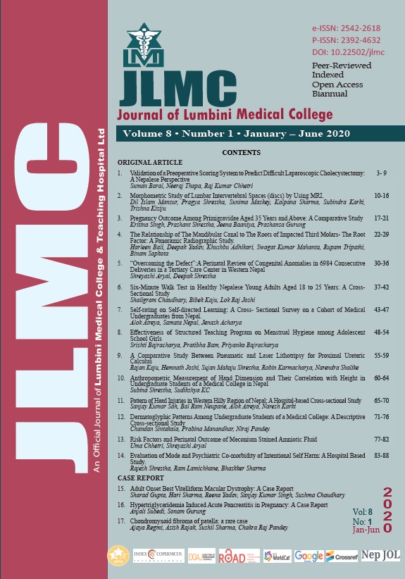
Morphometric Study of Lumbar Intervertebral Spaces (discs) by Using MRI
Keywords:
Intervertebral disc, lumbar vertebrae, MRI scanAbstract
Introduction: The radiological space between two vertebrae is known as intervertebral space (height) which corresponds to the thickness of the intervertebral disc. Lumbar intervertebral disc is the most important structure which maintains the spinal function. An early diagnosis of pathological changes in disc has clinical significance. Hence the study aimed to determine normal height of the intervertebral disc space and effect of aging.
Methods: It was a cross-sectional analytical study performed on 106 images of MRI scans of lumbar region. Dimensions of lumbar intervertebral spaces (discs) such as the anterior, middle, posterior intervertebral space height were measured in millimeter.
Results: The mean anterior intervertebral space height was gradually increased from L1-L2 level (6.91 mm) to L5-S1 level (13.55 mm). The middle intervertebral space height increased from L1-L2 level (7.89 mm) to L4-L5 level (11.96 mm) whereas at L5-S1 level, there was a decrease (11.10 mm). Similarly, the posterior intervertebral space height showed an increment from L1-L2 level (5.52 mm) to L4-L5 level (8.09 mm) except at L5-S1 level, where it was decreased (6.94 mm). All mean values were found to be higher in males than in females except posterior intervertebral space height. The height of disc was increased up to third or fourth decade followed by a decrease.
Conclusion: Knowing the normal lumbar intervertebral space height could be helpful for clinicians to diagnose and plan for proper treatment. It may also help to generate baseline data and to produce proper devices for Nepalese population.
Downloads
Downloads
Published
How to Cite
Issue
Section
License
Copyright (c) 2020 Dil Islam Mansur, Pragya Shrestha, Sunima Maskey, Kalpana Sharma, Subindra Karki, Trishna Kisiju

This work is licensed under a Creative Commons Attribution 4.0 International License.
The Journal of Lumbini Medical College (JLMC) publishes open access articles under the terms of the Creative Commons Attribution(CC BY) License which permits use, distribution and reproduction in any medium, provided the original work is properly cited.JLMC requires an exclusive licence allowing to publish the article in print and online.
The corresponding author should read and agree to the following statement before submission of the manuscript for publication,
License agreement
In submitting an article to Journal of Lumbini Medical College (JLMC) I certify that:
- I am authorized by my co-authors to enter into these arrangements.
- I warrant, on behalf of myself and my co-authors, that:
- the article is original, has not been formally published in any other peer-reviewed journal, is not under consideration by any other journal and does not infringe any existing copyright or any other third party rights;
- I am/we are the sole author(s) of the article and have full authority to enter into this agreement and in granting rights to JLMC are not in breach of any other obligation;
- the article contains nothing that is unlawful, libellous, or which would, if published, constitute a breach of contract or of confidence or of commitment given to secrecy;
- I/we have taken due care to ensure the integrity of the article. To my/our - and currently accepted scientific - knowledge all statements contained in it purporting to be facts are true and any formula or instruction contained in the article will not, if followed accurately, cause any injury, illness or damage to the user.
- I, and all co-authors, agree that the article, if editorially accepted for publication, shall be licensed under the Creative Commons Attribution License 4.0. If the law requires that the article be published in the public domain, I/we will notify JLMC at the time of submission, and in such cases the article shall be released under the Creative Commons 1.0 Public Domain Dedication waiver. For the avoidance of doubt it is stated that sections 1 and 2 of this license agreement shall apply and prevail regardless of whether the article is published under Creative Commons Attribution License 4.0 or the Creative Commons 1.0 Public Domain Dedication waiver.
- I, and all co-authors, agree that, if the article is editorially accepted for publication in JLMC, data included in the article shall be made available under the Creative Commons 1.0 Public Domain Dedication waiver, unless otherwise stated. For the avoidance of doubt it is stated that sections 1, 2, and 3 of this license agreement shall apply and prevail.
Please visit Creative Commons web page for details of the terms.


